
B-Scan ultrasonography
What is ocular ultrasound?
The ocular ultrasound uses high frequency sound waves to highlight the tissues around and inside the eye. The generated image in the classic ocular ultrasound is two dimensional and black and white. It is a painless, non-invasive procedure which lasts less than five minutes.
B-Scan Ultrasound
How is the ocular ultrasound performed?
The ultrasonography generates ultrasounds through a small stylet. The stylet is placed on the surface of the eyelids with the eyes closed. The patient feels only the gel that is used on the spot where the stylet will contact the surface of the eyelids. Then the reflected ultrasounds are collected and processed to form the image of the area that is examined.
In some special applications (UBM ultrasound) the stylet rests directly on the eye, after the application of a local anesthetic.
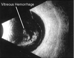
Ultrasound imaging
When is an ultrasound performed?
The ultrasound is performed in addition to the ocular examination to examine the tissues on the interior of the eye when a fundoscopy is not possible, e.g. in vitreous hemorrhage or very dense cataract.
Other applications of the ocular ultrasound are:
- The diagnosis and monitoring of intraocular formations-tumors
- The study of optic nerve anomalies (edema, Drusen)
- The detection of foreign bodies or dislocated lenses.
- The study of intraocular inflammations
- The study of extraocular muscles and of the orbital tissues
- The study of glaucoma