OCT Angiography – Angiovue
EYE DAY CLINIC was the first clinic to adopt the new, innovative imaging technique, called OCT Angiography. It is a method that provides high contrast images of the blood flow in the retinal vessels, which in the past was only possible with the classic angiography procedures that included infusion of dye in the eye: fluorescein angiography using fluorescein and indocyanine angiography.
Pioneering OCT- dye-free angiography
With the new imaging method, OCT-angiography, the imaging of the entire retinal vasculature as well as of the choroid capillaries is achieved through a procedure that is performed QUICKLY – PAINLESSLY – NON-INVASIVELY and without fear of complications/ side effects from the use of contrast media.
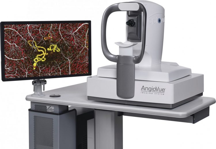
This examination is performed with Avanti XR, which is the top OCT model by Optovue and offers high resolution, quick scans (70.000 a-scans/sec), widefield scans and 3D reconstructions.
SSADA technology is used for tomography analysis, to achieve the detailed imaging of movable structures, i.e. of the blood cells inside blood vessels is produced.
Apart from the obvious advantages over classic fluorescein angiography, OCT-angiography also offers the following:
- Per layer imaging of the vasculature, transforming angiography from a two-dimensional to a three-dimensional examination.
- Detailed vessel imaging, without being obstructed from phenomena such as leakage, pooling, staining and hemorrhages which in fluorescein angiography may hide information.
- Clear imaging of the capillary network
- Imaging of the vascular network of the optic disc that offers significant information for glaucoma patients.
- Ability to frequently repeat examinations in patients with vascular diseases of the retina, who are submitted to treatment with intravitreal injections to determine the best possible treatment regimen.