OCT Angiography (without contrast media)
In this examination, the flow of the retinal and choroidal vessels is recorded without an intravenous administration of contrast dye. This is achieved by the AVANTI XR optic tomography, which is available for the first time in Greece at our fundus department.
Offering 70,000 A scans/ second, the SD-OCT AVANTI XR is the fastest and most advanced Spectral Domain OCT and the only device which can reproduce 3D retinal angiography images using SSADA technology.
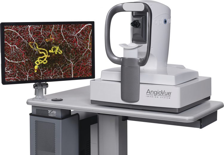
AngioVue with Angio Scan
In contrast to classic fluorescein angiography, where only the depiction of the outer capillary network is possible, dye-free OCT – angiography can depict the flow of both the inner and outer retinal capillaries.
Furthermore, the various layers of the retina and the inner choroid as well as their respective perfusion can be depicted individually, providing important information on a variety of fundus-related conditions.
Dye-free OCT angiography has many and significant advantages over classic fluorescein angiography:
- As a dye-free, non-invasive examination, it is harmless for the patient as, unlike fluorescein angiography, there is no risk of allergic reaction.
- Furthermore, it can be performed on patients with renal disorders or cardiovascular diseases.
- It does not require the presence of an anesthesiologist
- It does not require mydriasis
OCT – angiography is already being applied to the diagnosis, study and monitoring of various choroidal retinal conditions, such as:
- Age-related Macular Degeneration
- Diabetic retinopathy and diabetic macular edema
- Retinal vein occlusion
- Macular telangiectasias
- Optic nerve disorders