
Optical Coherence Tomography (OCT)
Optical Coherence Tomography (along with angiography) is the most significant and frequently used imaging examination of the retina and, more specifically, of the macula.
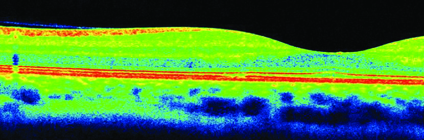
OCT examination image
It is a non-invasive and completely painless procedure, as a beam of light is used, which hits the retina and produces a pseudo-anatomical image of the macula.
The state-of-the-art Spectral Domain Optical Coherence Tomography systems offer high resolution imaging and can depict in greater detail the layers of the retina and of the underlying choroid, providing important information for many conditions affecting the back of the eye.

OCT examination image
The examination lasts a few minutes and can usually be performed without dilating the pupil.
OCT is now a necessary tool for the diagnosis and monitoring of the progress of many retinal conditions, such as age-related macular degeneration, macular edema associated with diabetic retinopathy or vein occlusions, the central serous chorioretinopathy, the macular hole, the epiretinal membrane, the vitreomacular tractions, etc.
OCT is also very important in the diagnosis and monitoring of glaucoma. It allows the mapping of the optic nerve and of the nerve fibers, the early diagnosis of glaucoma (before impairment of the visual field) and the recording of any changes in the progress of the disease.
Moreover, OCT has an application in the study of the anterior segment of the eye, the cornea and the angle of the anterior chamber.